Showing posts with label NFE2. Show all posts

Monday, September 09, 2013

Intra- and interchromosomal interactions of point mutations occurring in the vicinity of the normal 5-and 3 ends via low and high O(2)-affinities on the beta-globin complex.

Beta-globin (HBB) locus: 11p15.4  [§§; †, ‡-(HbS)] intra- and interchromosomal interactions with element in the beta-globin HBB is one of the 2 types of an asymmetric purine : pyrimidine sequences in beta-thalassemia patients (Hydroxyurea) and normal (nonthalassemic) individuals from the standard neutral – model, to any one or more of 200 different mutations (unstable free globin chain subunits), a heterotetramer subunits assembly composed of ‡ two α-hemoglobin chains and two β-hemoglobin chains. In adult (Hb) hemoglobin, the IVS-2-intron“‘ promoter a coregulator of the GATA1 can serve a similar function as NF-E2 here; chromatinized minichromosome associations in erythroid cells. These data indicate (CTCF-CCCTC binding factor, interactions affects spatial distances) observations that favor EKLF’s red cell (RBC) activators erythroid specificity.  A self-organizing process, proposed role activates an adjacent promoter as both (human fetal (gamma)-to adult (beta)-globin) are important, however not sufficient (basal) stabilizing interactions,  -both were in cis and in trans distinct from alpha-globin mRNA, the 2 types of polypeptide chains interrupted by 2 intervening sequences the so-called** “switch“* region (that is, gamma—-beta -the average zeta potential, of externalized phosphatidylserine minimal for zeta-globin HBZ  dissociation constants (fast or slow* moving), to an embryonic alpha-like hemoglobin),.  Gene-proximal acting cis-regulatory DNA elements (chromatin) are maintained that contain informative mutations ‘one’ on the 3-prime side of the beta-globin gene ‘and a leftward’ rate of neutral mutation (in the 5-prime direction) the centromere (beta-globin within the chromatin domain) which contains a ‘hotspot‘ (mutations causing diseases at HRAS1, D11S at one or more 11p15.5 loci in the HBB region from D11S and IGF2: INS are systems found to be dependent on EKLF ) for recombination in the HBB gene region 3-prime to the beta-globin gene (β-thal) mutations (led to DAPI lentiviral vectors (LVs) particles expression-cassette detection: genetic diagnosis (PGD) Preimplantation. And targeted integration of the adeno-associated virus (AAV).) at 5-prime splice sites (A gamma-) globin (HBG1) are held to be responsible for human genetic disease of fetal ‘Aγ and Gγ’ hemoglobin (HPFH/beta o-tha the BCL11A variant is associated with the same variable HbF) by (tagging with GFP) a single initial deletion followed by spread of the mutation, naturally occurring allele-(Hardy-Weinberg principle),  locus with two alleles denoted, and a second abnormal allele of an HBB mutation (e.g.,  the sickle-cell haemoglobin gene Hb S, a naturally occurring mutant Hb C, β-thalassemia), with subsequent crossovers between the 5-and 3-prime and gene conversion and the creation of 2 others (e.g., Comparison‘s of the normal 5-and 3 ends, the processive region 3′ to the 3′ UTR messenger mRNP complexes ribonucleoprotein breakpoint via mutations or HS deletions (β-globin HS5 or 3′HS1) that contributes to the abnormal expression, or as RNA stability, maturation and transcriptional termination) for recombination (crossing-over or gene conversion) both in cis and in trans intra- and interchromosomal interactions of point mutations occurring in the vicinity of the beta-globin complex,  in cis to the gene mutations, were physically intact. SATB1 takes part in affecting the HBB higher order chromatin structure Matrix attachment regions (MARs) within the locus control region (LCR located at the 5′ end, flanked by AAV),  the HS2 and 3′HS1 active chromatin hub (ACH), remote 5-prime element genes (a member of the HMGB-2 high-mobility group protein 2 family) in cis to the deletion a single initial deletion is the beta zero type of  a coexisting thalassemia component and if so, if it is α-thalassemia or Beta (gamma-beta-Thalassaemia and (SCD-Hemoglobin) Hb SS anemia, sickle cell disease) and malaria  has some protective effect from increased risk of G6PD deficiency, with beta-globin co-inheritance a fetal adult gene as a cofactor involving the first non-coding near the 5-prime end of 3 exons  plus a single pseudogene termed psi beta 1 ( epsilon, beta and gamma are complementary to the structure of genes is coincidental of site mutants that are turned on and off ( H3 acetylation-(H4/R3* in the R state having T/R** low and high O(2)-affinities)-K4 demethylation) the mechanism is more complex as development proceeds) the Dominant Control Region (DCR) and introns“‘ 1-5 both single nucleotide“‘ substitutions of the beta-globin gene to the deletion ‘in cis‘ a region designated LCRB, locus control region. (INS) the insulin gene was also mapped to this same region.


hbb
(1)  the "hinge region" of the alpha 1 beta 2 interface PMID: 1567857 were partitioned into components of ( PDB:1J7Y_colored in reds is Hb-alpha ) SNP PDB:1IRD HBA1 and 2 structure rearrangement,  the interface from the mutation site is site (B) about protein sequence 4L7Y-B alpha and D-beta: Resultsare for rs33930165 on Reference Sequence: NP_000509.1 [PMID: 22028795] attainment number P68871 verified by refinement of the a entire  molecule was confined to residues at the central cavity close to the 2,3-DPG found in the NP_000509.1 hemoglobin (PDB: 4L7Y) subunit beta. 1J7Y_Reds Hb-alpha,_Blues Hb-beta. With The effect of mutagenesis on O(2), CO,                   and NO binding to mutants 1J7Y HBB.H116R_D test Disease Gene: HBB  protein/NP_000509.1structure arrangement. The alpha (HBA) and beta (HBB) loci determine the structure resolution analysis reported here implies...  the structure of genes is
coincidental of site mutants that are turned on and off ( H3 acetylation-(H4/R3* in the R state having T/R** low and high O(2)-affinities)-K4demethylation) the mechanism is  more complex as development proceeds) e.g.  not present in the final mature HBB gene product.

hbb


(2)  Behaviour of a natural haemoglobin and a mutant variant in the central cavity close to the 2,3-diphosphoglycerate pocket  4L7Y-D a band migrating in the Hb F_ a solvation band-position-PDB: rasmol_php (DiseaseE6K_33930165_F_[solvent- is nonbonded spheres on 4L7Y-D Hb-beta Red fig. (1)) and its reactions with 2,3-DPG and inositol hexaphosphate-PMID: 6526653: accounts for the reduced oxygen affinity of haemoglobin;  by the oppositely charged side-chains residue that project into or are missing in the heme pocket, and result in a thalassemic and/or hemolytic -like phenotype the result of decreased alpha 1 beta 1 interactions.
hbb
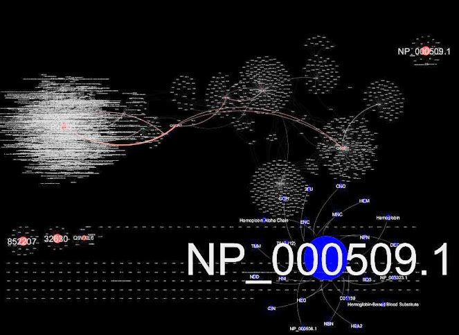
HBB Network visualized with Cytoscape. The inverse of the inverse not inferable from Figure (4) overlaps the hinge region for exon selection 3'5'duplications. pubmed/21269460 [#35]
hbb

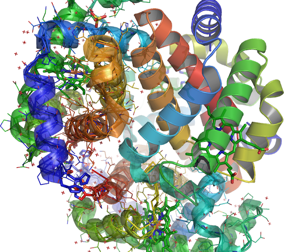
 (3) 4L7Y-B inhibits the rate of ligand binding HIS'147 the native imidazole side chain is 4L7Y-D modification at each site is a function of the position of these 2 hemoglobin alpha and beta introns the electrostatic attraction or repulsion by the oppositely charged side-chains therefore the efficiencies of intron 1, PMID: 6599969 and intron 2, PMID: 16184579 are unaffected residue near the 3' end (Blue color) (4L7Y_B/B/LEU'3/CA) of the intron on a mechanism that measures the distance, the first intron might facilitate splicing (aligned as B-D, B-D) of the second intron (Orange) 4L7Y and desease HBB locus gene in which intron 1 PMID: 18266765 accommodates the 5' end (Orange). Introns are not present in the final HBB gene product mature RNA with SNP: rs33949930,                   amplified from exon (Blue) 1 + 2 (PMID: 8226093) of the beta-globin gene: NG_000007.3, (a neutral mutation [ SNP: rs33949930                   Position 70599 http://tinyurl.com/nhut5yf]). Present in SNP to nucleotide allele T.
hbb
(4)  Correlated inversely. The intron is linked both in the intron-exon sequence and nearer the (Blue) 3' end (an adaptation to endurance PMID: 16990440 ) of the intron upstream from the 3' terminus to the 3'-side of the beta-globin gene PMID: 478302 of the intron (Orange) on 4L7Y-B beta-globin gene should remain active together with all other (PMID: 11559912 alleles) forms of the same HBB gene multiallelic loci  PMID: 15315794 involved in beta-thalassemia along with the unrecognized allelism found in  PDB:1IRD among a new neutral mutation. V2E, A, G, L, SNP 33949930 (hydrophobic interaction decreased; hbb hbb )  the single nucleotide polymorphisms NP_000509. The remaining 95% of the SNPs for prediction in which a variant could be detected, would have been sufficient in these cartoons, however may be misleading.  These results suggest that e.g. the introns (PMID: 11860449) or the entire Hb-beta locus may be  missing in beta(0) or be impeded ( O(2)-affinities) in Hb SS anemia beta-thalassemia and if so, α-thalassemia or Beta (gamma-beta-Thalassaemia and (Sickle Cell SCD-Hemoglobin)  Hb SS anemia, sickle cell disease.

Wednesday, May 25, 2011

The amino acid human KIAA0132 protein central BTB/POZ domain double glycine repeat (DGR), Keap1.


 1.35 angstrom DGR structure of the Kelch domain of Keap1
1.35 angstrom DGR'' structure of the Kelch domain of Keap1
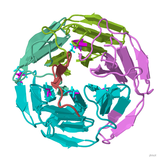
 Kelch beta-propeller lining the central channel of the propeller, interacting with residues in loops between strands of the the six-bladed [beta] 5-exon structure making contacts with conserved residues in the Kelch repeat Both the N- and C-termini of the domain are located in gray monochrome blade. PDB Structure: 1ZGK
Kelch-like Ech-associated protein-1, or Keap1 locus: 19p13.2 [§§], encoding phase 2 detoxifying enzymes and antioxidant stress proteins, is identical to the amino acid human KIAA0132 protein central BTB/POZ domain double glycine repeat (DGR), or Kelch, module. Keap1 interact with the Neh2 domain of Nrf2. The crystal structure of the 6-bladed Kelch repeat contain the DLG and ETGE motif of Nrf2 bound the beta propeller of Keap1 at the entrance of the central cavity on the bottom side targeted PGAM5  to the outer membrane of mitochondria binds to the substrate binding pocket, and proper association of Keap1 with Nrf2; Keap1 acts upstream of Nrf2 in the cellular response. Defects of Keap1 activity transactivates the expression of several dozen cytoprotective genes  (The fetal Alz-50 reactive clone 1 (FAC1) protein and Sarcosine-KBTBD10, are ubiquitinated by a Cul3-dependent complex)  that enhance cell survival for their survival against apoptosis when Nrf2 is released from Keap1 dependent degredation functions as a sensor of oxidative stress and cellular redox homeostasis imbalance. Upon exposure to redox electrophiles a putative ARE in the GI-GPx promoter glutathione peroxidase 2 (gastrointestinal), quinone-induced oxidative stress, sulforaphane (SFN) and curcumin  xenobiotic (XRE) stresses may be therapeutic neither disrupts association between Keap1 and Nrf2, and by reversal of these effects whose mutational status is associated with inactivating a mutation or two of the 6-bladed Kelch repeat compatible with proliferation preventing Nrf2 activation of antioxidant response element (ARE) mediated gene expression in the cytosol located in the cytoplasm of the cell bound to and  is repressed by the cysteine-rich Keap1 that translocate to the nucleus. Keap1 serves as a redox-regulated substrate adaptor protein that, an antioxidant response and stress-responsive genes imbalance lead to alteration of the Keap1-Cul3 [cullin 3] interaction the N-terminal regions hypoxic and unstable oxygenation microenvironment of a tumor, CAND1-cullin-associated and neddylation-mediated substrate adaptor recycling is required for efficient repression of Nrf2.


Sunday, May 22, 2011

Cis-elements as Maf-Maf homodimers/Maf-Nrf2 heterodimers and its negative regulators like p45 NF-E2 in the Nrf2/Keap1 system.


 Structure of the Keap1:Nrf2 interface.
 The Nrf2 peptide contains two short antiparallel beta-strands connected by two overlapping type I beta-turns stabilized by the aspartate and threonine residues.  
Structure of the Keap1:Nrf2 interface
Transcript Variant: NRF1. This variant (1) represents the longest transcript and encodes the longest isoform PDB Structure: 2FLU
NRF2 is the primary regulator of this endogenous antioxidant response, (nuclear factor erythroid 2-related factor 2) is located on 2q31: [§§] encoding NFE2 (an essential regulator of megakaryopoiesis), NFE2L1, and NFE2L2 basic-leucine zipper (bZIP) transcription factors, which play roles in oxidative and the e.g. (StRE/ARE) presumed natural antioxidants sulforaphane (SFN) and curcumin  xenobiotic (XRE) stresses may be therapeutic for cholestasis preventing carcinogenesis. However, the molecular mechanism of this regulation remains resolved in the six-bladed beta propeller crystal structure of the Kelch domains, the Cap-N-Collar (CNC) transcription factor family (BACH1) and its negative regulators like p45 NF-E2 in the Nrf2/Keap1 system. Nrf2 activation is mainly cytoprotective.  Nuclear accumulation of erythroid derived 2, like (Nrf2) increase the expression of the antioxidant HMOX1, and the expression of stress-responsive genes responsible for reactive oxygen species, (ROS) promote megakaryopoiesis elimination during maturation where HIF-1 is primarily induced in  the hypoxic and unstable oxygenation microenvironment of a tumor in several human cancers. Nrf2 dominant-negative mutant with the NQO1/NQO2 gene nuclear proteins a prototypical Nrf2 target cytosolic protein gene that catalyzes the metabolic reduction of quinones bind to the six putative ARE (antioxidant response cis-elements) as Maf-Maf homodimers (the term "Maf" is derived from MusculoAponeurotic-Fibrosarcoma virus) and Maf-Nrf2 heterodimers hMaf heterodimerizes specifically with Nrf1 and Nrf2, human BSEP (bile salt export pump) and the conjugate efflux pump (dietary chemopreventive agent sulforaphane (SFN) similar oxidized low-density lipoprotein is attributed to toxicity by siRNA-Nrf2)  reveals two similar musculo-aponeurotic fibrosacroma (Maf) recognition elements (MAREs). Nrf2 is released from Keap1, and translocates to the nucleus, Keap1 (Kelch-like ECH-associated protein 1) is the major upstream regulator of Nrf1, a BTB-Kelch substrate adaptor protein, through cis-active sequences (TXAS gene utilizes the same cis-acting element) known as antioxidant response element ARE is controlled by the NES nuclear export function Keap1 counteracts, that translocates in the nucleus once, in the cytoplasm ubiquitination targets Nrf2 for degradation (CRIF1, unlike KEAP1, both N- and C-terminal regions), and hence typifying oxidative stress-mediated apoptosis that lead to alteration of the Keap1-Cul3 [cullin 3] interaction (cytosolic and mitochondrial glutathione (GSTs) and protein-thiol redox imbalance), can no longer serve to target Nrf2 for ubiquitination, though it retains its affinity for Nrf2. The INrf2 (Keap1)/Cul3-Rbx1 [ring-box 1, E3 ubiquitin protein ligase] complex constantly degrades Nrf2 under normal conditions. ENC1-mediated  (ectodermal-neural cortex 1) down-regulation of Nrf2 was independent of Keap1, ENC1 is a BTB-Kelch protein and belongs to the same family as Keap1. Whereby Nrf2 activity is beneficial in non-malignant cells in  human neuroblastoma cells it may provide a advantage in ECs, containing several Nrf2 target genes (NQO1 and HO-1), induces MRP1 (Multidrug-resistant proteins) upregulation, leading to overexpressed its target Trx-1 [Thioredoxin] such as in human breast cancer cells for their survival against chemotherapeutic agents one of the at least two trans-acting transcription factors Polyamine-modulated factor 1 (PMF-1) binds to NF-E2 related factor-2f  that, Spermidine/spermine N(1)-acetyltransferase SSAT is regulated by.

Thursday, September 03, 2009

v-MAF as Compared with Microsomesis by de-repressing by binding Bach1's BTB and CNC homology

Group VI: RNA reverse transcribing viruses; Maf
hMAF/CNC-bZip heterodimers lacks any recognizable activation domain. NRF3 is able to dimerize with MAFG related coding for a small MAF transcription factor the v-maf ontogeny [Gene-Ontology-based functional annotations] regulated through nuclear factor erythroid-2 (NFE2) elements derived from the globin locus control region: [§§; 17q25] containing a ubiquitous small Maf protein (MafF, MafG, or MafK), cDNA encoding human MAFG, in megakaryocytic culture MAFK and MAFG mRNAs were induced similarly to BACH1 mislocalization locus 7p22 in erythroid culture, is expressed in a wide array of tissues and cell lines, the human MAFG contains at least 3 exons, which are separated by small introns. NRF3 transcripts are present in placental chorionic villi from at least week 12 of gestation on through term. Basic leucine zipper (bZip) domain in common and are subdivided into large and small Maf proteins.
MAF From the following article: Cancer chemoprevention with dietary phytochemicals