Showing posts with label G6PD. Show all posts

Saturday, April 09, 2016

TRIOSEPHOSPHATE ISOMERASE (TPI) A DIMERIC GLYCOLYTIC ENZYME AS A MODEL OF TIM-BARREL ACTIVE-SITE STRUCTURAL AND CHEMICAL ASPECTS IN THE MONOMER LOOP REGION'S REVERSIBLE CATALYTIC REACTION.


Triosephosphate isomerase (TPI, EC 5.3.1.1) (§, ‡) is essential to glycolysis, catalyzes the fifth step in the glycolysis pathway the reversible conversion of dihydroxyacetone phosphate (DHAP) into glyceraldehyde-3-phosphate. TPI is a homodimer formed by two identical dimeric molecules of a single structural locus : 12p13.31. TPI has only 1 functional gene with a molecular mass of 29 kDa, that after refinement are products of a distinct single structural locus. The variant phenotype of identical subunits are expressed in both red cells and circulating lymphocytes, catalyzing the interconversion of one of the two products breakdown by reversible conversion. The TPI substrate by deprotonation the transition state reaction of dihydroxyacetone phosphate (DHAP) substrate yields one product of the glycolytic pathway, is a trend* (Kcat) that persists creating the initial complex microcompartmentation of TPI to give (G3P) glyceraldehyde-3-phosphate which seems to be the isomerase* activity, release is slower than its conversion to DHAP in normal and TPI deficient cells. TIM with its natural substrates has not been (•) crystalized**. TPI is a dimeric enzyme and contains 7 exons interrupted by six introns.

monomers The crystallographic structure of (HsTPI) human triosephosphate isomerase PDB:1HTI is one dimer per asymmetric unit subunit 1 and subunit 2 are in the open and closed conformations in the 3-dimensional asymmetric space group P 2(1) which is specific to the Monoclinic with minimization on the entire structure in the presence of substrate analogues and its surrounding residues supporting possible regions targeted for drug design.


TPI deficiency (TPID) a disorder of glycolysis, occurring in haplotypes of specific alleles heterogeneous to clinical TPI-deficiency, with a rare homozygous deficiency the resulting genetic defect is the cause of a null variant incompatible with life by abnormally high levels of DHAP which degrades spontaneously into the toxic (MG) methylglyoxal, due to deamidation of asparagine (Asn15-71) to form aspartic and glutamic acid. Loop 6 plays a role in preventing the breakdown yield of methylglyoxal (fMG) one of the of the three products of enzyme-bound enediol(ate) phosphate, towards elimination of (fMG) inorganic phosphate. TPI deficiency is due to the common aberrant dimerization (or the dissociation into inactive monomers) of mutation TPI 1591C, encoding a Glu104-to-Asp (glutamate-to-aspartate) substitution in the TPI variant found in cases of hemolytic anemia coupled with neurodegeneration, the Glu104-to-Asp substitution is the most common disease allele inherited, when compared to wild-type TPI's three (residues from the same subunit) similar but not identical interactions between the inhibitor and catalytic residues, Glu 167 (or 165) forms a stable dimer and provides the rationale for production of structurally normal enzyme in humans, the E104D mutation, provides the amyloid-resistant structure of human triosephosphate isomerase (HsTPI). Water-protein molecules join two catalytically active monomers which is only in its dimeric form, as monomers of TIM are not functional. Within a hydrophobic catalytic pocket of the native enzymes the binding and catalysis of TPIs in hemolysates, bind to the red cell membrane. Molecular modeling using the human crystal structure of TPI was performed to determine how these mutations could affect enzyme structure and function. The Amyloid secondary structure autoepitopes antigen-driven mechanism works toward recovery of the anti-triosephosphate isomerase mutant TPI peptide** antigens. This is the scheme that allows function-enhancing stability most significantly, the catalysis for deprotonation of DHAP or vice-versa GAP substrates of the TIM-barrel relative to TPI toward turnover of two-part substrate glycolaldehyde / phosphite dianion {GA + HPO32* the transition state for this enolising enzyme substrate pieces.} Km/obsd* group of the whole GAP substrate and H95 (loop 4) is also optimal for small mutational changes in or reflects its compatibility with amino acid residues which stabilizes the enediolate intermediate (GA/HPO) activity from change in the products scheme (a proton transfer mechanism) DHAP/G3P or interconversion of these intermediates.

dhap-g3p

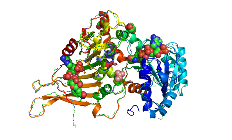
Closed (activated for catalysis) of optimal WT (TPI) molecular modeling PDB 1HTI_B using the human crystal structure of TPI human triosephosphate isomerase (HsTPI) conformation 1hti_b, calculated to the incidence residue Water-protein molecules and the protein cage that interacts within a hydrophobic catalytic pocket isolated and examined which coded for human triose-phosphate isomerase. [EC: 5.3.1.1]….


The active flexible site loop must open before product release, unliganded in trypanosomal Tb-TIM glycerol phosphate ester to liganded Glu167 in the catalytic cycle and the enzymes substrate transition state between open and closed to protect the substrate for the turnover of DHAP and G3P (GAP) the natural substrates, and inhibiting the formation of a toxic by-product in the absence of this equilibration reactions between dihydroxyacetone phosphate and glyceraldehyde 3-phosphate (G3P) enzymes by mutations that impair biosynthesis transforming competent cells, in the presence of an auxotrophic effect with these differences generated for an inability of the host organism to synthesize an essential compound during glycolysis in Tb-TIM. Trypanosomal-TIM is a glycolytic enzyme essential for the parasite survival that causes Chagas* disease, in this study G. bellum from the genus related Geraniaceae and its phenolic compound are leads which generates an unstable epimer of an enzyme Geranin A-containing changes resulting from ligand adducts in the active site to capture in addition a source of frustration that becomes more favourable. Glycolaldehyde (GA) the simplest sugar-related molecules uptake of a proton by Glu167 preserves the small effect for inhibition by PGA (transition-state analog) relative to substrate, G3P produces a triosephosphate isomerase with wild-type activity, loop 6 adopts the "closed" desolvated  (+) conformation to facilitate completion of catalysis by the formation of the › Michaelis-Menten complex (on the ‹ micros-ms › time scale) utilization yields further corrected calculations with corresponding (slower Kcat) motional rates* Km. Increase's are discussed in the context of the significance (Enzyme kinetics\Kcat) and may be estimated where the 'single-substrate' is locked in a protein cage probably  because of an active reaction site (loop 6) movement to the transition state for deprotonation; which are the on-average opened (substrate binding and release) and closed (activated for catalysis) of both monomers optimal WT (wild type) TIM conformations. Lys-12 ‹ is expected to interact with both centers, where the enediol intermediate along with the catalytic glutamate base and histidine-95 the catalytic electrophile stabalizes the reversible reaction intermediate that polarizes the substrate DHAP in the Michaelis complex. Interconversion spans the C-terminal end of the eight β-strands. For catalysis to occur likley a low pKa value transition from DHAP - for the enolase reaction enzyme enhancement 'relative to the nonenzymatic reaction - (Bound PGH - phosphoglycolohydroxamate mimics the (closed form) negative polarization (•) charge••, while PGA (2-phosphoglycolate) the positively charged residues in the two active conformation sites.) is similar for the two conformers' in the closed conformation, on ligand binding interacting with the reactive end's (β) the deprotonated substrate-bound structures to be protonated by a single-base (Glu-165) proton transfer^ mechanism.

philo

Structure of human triose phosphate isomerase at the positions of introns in homologous TPI genes from a number of phylogenetically diverse species. The introns motif are identified as calculated in phylogeny.
Phylogenetic trees constructed on the basis of sequence comparisons for triosephosphate isomerases analysis, TIM sequences were constructed based phylogeny with similarity, to those adopting the same structural fold of interest from different species for the taxonomic groups and the K13M mutations involvement in the human triosephosphate isomerase gene family...


Interactions in the loop regions combine the effects of His95 and Lys13 for Glu165 (loop 4, 1, and 6) the three crucial catalytic residues in triose phosphate isomerase, all form the enediol intermediate necessary for the interconversion reaction catalyzed by TIM resulting in the natural substrates G3P formation. The introns motif are identified as calculated in phylogenic motifs. Poorly conserved residues as targets for specific•• drug design are expected when compared to (TPI) Triosephosphate isomerase (•). Catalytic residues of the phylogenetic relationship pathways obtained by sequence based methods of specific key amino acids can than be calculated to the incidence residues and other TIMs which may influence the (human) HsTPI equilibrium.


Sunday, June 14, 2015

CHANGES IN GLUTATHIONE AND GLUTATHIONE REDUCTASE POSITIONING GLUTATHIONE-S-TRANSFERASE AS A FUNCTION OF CELL CONCENTRATION WITH ENZYME ACTIVITIES FOUND TO INFLUENCE BEHAVIOR.

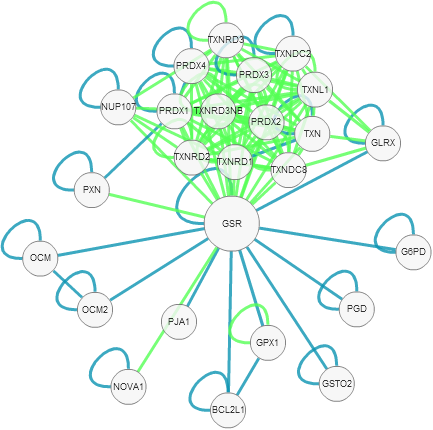
Glutathione reductase (GSR, GR) locus in the chromosomal region 8p21.1, (EC 1.8.1.7)-(§, ‡) is a protein-S-glutathionylation, as a (human) Mitochondrial localization of hGSR and its associated enzymes cellular thiol/disulfides S-Glutathione reductase (GSR) which is the importance of significance in reversible thiol modifications which  regenerates reduced glutathione (GSH) and GSSG to the reduced form found in the obvious structural properties of glutathione reductase. The redox regulating enzymes relationship with TTase (thioltransferase) activity with the ratio of the activities of G3PD, as the mechanism (of cellular repair) 'differs' (gssg-g6pg) according to the type of reducing glutathionylated mixed disulfide, including protein-S-S-glutathione (PSSG), GSR reduces (PSSG) modified by thiolation to a normal level in human lens epithelial (HLE) cells. This may have implications in stress- and aging-related pathologies in astrocytes and granule cells, demonstrated by comparable mitochondria/cytosolic concentrations of its thiol proteins, where a mitochondrial leader sequence (cDNA) is present in the gene structure of human GSR and may be the Cytoplasmic Isoform (derivative or inhibitor formed) of  mitochondrial dysfunction that contains the catalytic cysteine revealing a possible therapeutic strategy/target, also indicating transiently accumulated inhibitor proteins modified by thiolation (cysteine catalytic subunits) compounds that inhibit these (re)activation processes (hGSR) with its structure-based prosthetic group (FAD) cofactor is common because of the levels of cysteine available; are mitochondria/cytosolic concentrations that the Glutathione reductases reversible thiol modifications which catalyzes the reduction of GSSG to GSH the natural GR substrate is dependent on the NADPH:GS-SG ratio.
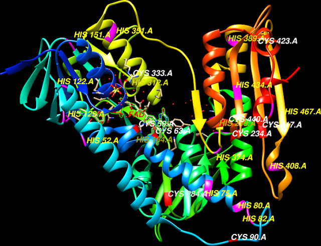
PDB Id: 3DK9 Cys58 and Cys63 represent the enzyme's results seen as the reductive (GSH) Cys-58 and oxidative (GSSG) Cys-63 is the relationship of these two enzymes, His467' is seen to interact with Cys63 more optimally and Cys-58 produces the second GSH intermediate molecule of the reaction is the reduced glutathione-to-oxidized glutathione ratio (GSH/GS-SG) when compared to the substrate free form correlated with (FAD) the flavin compounds, flow from NADPH to the substrate GSSG via flavin. The reducing equivalents needed for regeneration of GSH are provided by NADPH. The enzyme has affinity for flavin adenine dinucleotide (FAD) the prosthetic group of GR, and maintains high levels of reduced glutathione  (Cytoplasmic Isoform: Produced by alternative initiation of isoform Mitochondrial homodimer, derivative or inhibitor formed from the GSR Pyridine, dimerisation domain.) in the cytosol. Glutathione reductase (GR) plays a key role in maintaining either a thiol group or a nonprotein sulfhydryl group (NPS) form of GSH, and potential for thioredoxin and glutathione systems, as thioredoxin dose not require GSH and GR for catalytic activity. Glutathione reductase (GR) utilizes NADPH produced by G6PDH (glucose-6-phosphate dehydrogenase) enzyme activities, and enzyme glutathione reductase (GR) represents the erythrocyte glutathione-reducing system (GRS), of the GSH pathway to oxidation and inactivation in the activity of GSH peroxidase and GSH reductase. Expression of the regulatory subunit of gamma-glutamylcysteine synthetase/ligase (GCL) catalyzes the first and rate-limiting step in the production of the cellular (GSH) glutathione. Dietary riboflavin (Vitamin B2) intake produces its active essential coenzyme flavin forms, riboflavin mononucleotide (FMN) and flavin adenine dinucleotide (FAD) of glutathione reductase (GR), or the GR activity correlated with red-cell flavin compounds.When both GSSG and NADP(+) substrates and products are present, glutathione reductase (GR) is a enzyme required for the conversion in the presence and absence of flavin adenine dinucleotide (FAD), glutathione reductase (GR) is an obligatory FAD-containing homodimer. GSSG via glutathione reductase (GR) regenerates reduced glutathione which is essential for antioxidant defense. The flavoenzyme glutathione reductase (GR) reduces 'oxidized glutathione' (GSSG) back to GSH, also involving glutamate-cysteine ligase and modulatory (GCL)-can be upregulated ∉ as the cellular GSH system, indicating short-term and heritable tolerance of exposure to oxidative stress from/via numerous reporting ∈ mechanisms. NADPH is used by glutathione reductase for the reduction of oxidized glutathione (glutathione disulphide) GSSG to GSH-dependent peroxide metabolism. 4-Hydroxynonenal (HNE) is one of the major end products of lipid peroxidation which may lead to enhanced action of  the (GSR) oxygen radical, glutathione S-transferases (GSTs) are specifically suited to the detoxification and removal of 4-HNE (∋ or ∝) from cells which may provide a basis for selective cellular and/or subcellular distribution of mitochondrial and cytosolic to individual detoxifying gene inducer activities of glutathione reductase (GR), the cellular (GSH) glutathione. It was evident the enzyme glutathione reductase (GR) represents the erythrocyte glutathione-reducing system (GRS), of the GSH pathway to oxidation and the (∉ or ∝) inhibition constant for reversible inactivation in the activity of glutathione related antioxidant enzymes. And GSH reductase may be one of the factors that remained in focus that suggests its effects on the antioxidant system related to glutathione synthesis (GCL), degradation, and functions.

Biological Xenobiotics, Extracts, Applications of note In the presence of Glutathione reductase.:

Schisandrin (Schisandra chinensis), used in traditional Chinese medicine. PMID:21328628
Transketolase (TK) and transaldolase (TA)
Melatonin PMID:15571523, 19475625
Blackberry (Rubus sp.) cultivars, The 'Hull Thornless',  PMID:11087537
Glutathione dehydrogenase (ascorbate)-[dehydroascorbate reductase (DHAR), and glutathione reductase (GR). This enzyme participates in the glutathione metabolism the active metabolite of vitamin D3 increases glutathione levels.] PMID:11087537, 23770363
3H-1,2-dithiole-3-thione nutraceutical D3T potently induces the cellular GSH system, Anethole trithione is a drug used in the treatment of dry mouth, the Anethole trithione isomer is related to anethole (anise camphor) used as a flavoring substance. PMID:17206382*, 19408115,     19176875*, 15896789, 18408143*, Glutathione reductase
16946404*
Cassia fistula used in herbal medicine. PMID:19088944
Sanguinarine is extracted from some plants, including bloodroot and Mexican prickly poppy (Argemone mexicana) where argimone oil causes Epidemic dropsy. PMID:11260782
Vitamin E, PMID: 15672860
Tocotrienols are natural compounds members of the vitamin E family found in select vegetable oils are an essential nutrient for the body. PMID:21845802
Pyrrolizidine alkaloids are produced by plants as a defense mechanism against insect herbivores consumption of PAs is known as pyrrolizidine alkaloidosis. PMID:20144959
Apple extract (AE) PMID:20401791
Lipoic Acid an organic compound, forming a disulfide bond, available as a dietary supplement PMID:15246746, 21073761
Carnitine PMID:15246746, 10581232
Vitamin D upregulated expression of GCLC and GR. PMID:23770363
Vitamin D3_ PMID:12416023
Vitamin E_ PMID:10459841, 8360018, 18296478, 21845802, 15490422, 16885600, 7062348, 20729758, 21086752
Shidagonglao roots Mahonia fortunei (十大功劳 shi da gong lao) species contains the alkaloid berberine PMID:199382 18
Coenzyme Q10 (CoQ10) PMID:16621054
Trigonella foenum graecum seed powder (TSP) PMID:15026271
Boschniakia rossica, a ̱̱̱Traditional Chinese medicine. PMID:19352025
Aegle marmelos commonly known as bael is a species of tree. PMID:18830880
Scoparia dulcis A medicinal plant, dulcis. PMID:21905284
Fenugreek (Trigonella foenum-graecum)  is used as a herb. PMID:15026271
L-arginine (L-Arg) semiessential supplementation common natural amino acid. PMID:16038634
Hypericum perforatum (St. John's Wort) PMID:18754092
Urtica dioica often called common nettle PMID:12834006
Usnea longissima, a medicinal lichen. PMID:16169175
Capparis decidua, a fruting tree also used in folk medicine and herbalism. PMID:22272107
Indole-3-carbinol found at relatively high levels in cruciferous vegetables such as broccoli
PMID:9512722, 14512388
Ascorbate Vitamin C. PMID:14512388
Sulforaphane It is obtained from cruciferous vegetables such as broccoli. PMID:12628444, 18607771*, 22303412
Andrographis paniculata, may shorten the duration and lessen the symptoms of common cold. PMID:11507728
Vitamin B-1 (thiamine) PMID:1132146, 10450194, 21308351*, 11514662*, 1270885
Vitamin B2 (riboflavin) PMID: 5822598, 5550591, 1201246, 5794396, 237845, 3677785, 3582603, 12194936, 2721660, 1261528, 5721130, 14608016, 4400882, 7883462, 844948, 7337797, 5881,12641409, 4393763, 3497609, 16883966...(№ 1244, OMIM.138300)
Vitamin B-6 (Pyridoxine) PMID:2721660, 3582603, 10450194, 15490422, 1270885, 7417521, 7337797, 7814235
Vitamin B9 (Folic acid)  PMID: 844947, 1270885
Aspartate transaminase (AST) or glutamic oxaloacetic transaminase (GOT) catalyzes the interconversion of aspartate an important enzyme in amino acid metabolism. PMID:1132146, 10450194, 1253408
β-Carotene is a strongly colored red-orange pigment abundant in plants and fruits. PMID:19957244
3-Hydroxykynurenine (3OHKyn) a metabolite of tryptophan. PMID:11273669
Ajoene ((E,Z)-4,5,9-trithiadodeca-1,6,11-triene 9-oxide), a garlic-derived natural compound. PMID:9986706 PDB: 1BWC
Propolis a product made by bees. PMID:19394397
Resveratrol produced naturally by several plants PMID:12797471
 No CiTO relationships defined:
 http://vixra.org/abs/1506.0104
 http://www.citeulike.org/user/emissrto/article/13645622

Monday, December 30, 2013

G6PD, Exon 12 is an exonic splicing silencer containing/substituted define codon regions involved in the G6PD mRNA¹

G6PD (EC 1.1.1.49) glucose-6-phosphate dehydrogenase [§§; †, ‡], situated at Xq28 locus-coding region is the rate-limiting enzyme, of the (PPP) pentose phosphate pathway. G6PD deficiency  and its  X-linked gene mutations exons 2-13 (160 different mutations) are the most common inborn error of metabolism, in human red blood cell (RBC) enzymopathy, among humans. G6PD is divided into 12 segments and involves an exonic splicing enhancer (ESE) in exon 12 with 13exons and an intron present 5' UTR, proximal to the 5' bkp-breakpoint region. Intron comparisons from the second to the thirteenth exons of G6PD gene, 3′ UTR towards the 3' end of the gene to exon 1 located in 5' UTR G6PD is a region of deleted alleles (ASO-PCR) or G-6-PD the many population genetics variants/wild-type (160 different mutations and  300 G6PD variants) assuming that, at exon2 (2,3-BPG* levels) are hypothesized that G6PD partly 'overlaps' the IKBKG gene confined to the blood. The subunit (G6PD), consists of the biochemical-characteristics of 531 amino acids. This enzyme is the only process in mature red cells for NADPH-generation it involves oxidation of glucose as a » hexose « ( xenobiotic compounds) pathway ('naturally found in D-* and the unusual L- Monosaccharide forms or between 2,3-BPG*) pentose and hexose phosphates, an alternative to glycolysis, converts glucose in which ATP is produced' from the conversion of glucose-6-phosphate into ribulose 5-phosphate in liver cytosol in which a residue in the dimer interface (@ 37° C) structural G6PD is a NADP+ dependent. At the tetramer interface an Apoenzyme (PDB:2BH9), that stimulates G6PD to produce (reversible enzyme transketolase (TK) presence is necessary) more NADPH. Hemolytic crises or dysregulated NADPH oxidase located in the 3' dependent 5' UTR G6PD in humans determines the response, in which G6PD deficiency is prevalent with development of  chronic hemolytic «« anemia (CNSHA-HNSHA) associated with food-induced or a exogenous-agent and drug-induced º' ª hemolytic crises which led to the discovery of G6PD deficiency. Sulfatase  (STS, EC 3.1.6.2) catalyzes Phenyl-Piracetam [↩] it also stacks well  and involves the phosphoinositide 3-kinase (PI 3-kinase) pathway in the employed doses in related induction of certain enzyme (Glucose 6PD) synthesizing activities (glycolysis) five metabolite levels of  insulin signal transduction. These include, Sulforaphane  or broccoli-sprout extracts increased cell-lysate NAD(P)H:quinone oxidoreductase (NQO1) phase II activities (Tanshinone IIA⊕), administered to cells and  in human supplementation studies, were found to be in balance with green tea extract (GTE), (EGCG) epigallocatechin-3-gallate   to generate detoxifying reactions to hepatotoxicity (can be prevented by amalika, Emblica officinalis   which supports the chemopreventive action of the silymarin extract Silibinin , of the milk thistle) preventing nitric oxide-mediated lipid peroxidation (LPO) and antioxidant defense system (GSH) glutathione ( GSH-Px and GR) depletion, via an antioxidant response element (ARE ⊕) mechanism-based inhibitor, element (NRF2) regulates (ARE)-regulated genes. A lack of NQO1 protein predisposes cells to benzene toxicity and to various forms of leukemias and toward therapeutic modulation (Acetylcysteine  and acetaminophen) of pulmonary oxygen toxicity. G6PD-deficient variants is the result of  various enzymopathies (but not GPI-chronic hemolysis), that glucuronidated-bilirubin values (UGT1A1 genotype) tended to parallel, (CNSHA) hyperbilirubinemia with hemolytic anemias, single amino acid substitutions resulting in 'mutation of variants'. Or to inherited³ and acquired physiologic changes in red cell enzyme G6PD deficiency leading to favism ( an A- variant reaches the polymorphism level the commonest a Mediterranean form, other alleles A, A+, the primordial human type B cell and normal B+ and a rare B- phenotype are neutral. Malaria-infected human red cells possess at least two pathways (in a dimer -- tetramer equilibrium) where carbonic anhydrase (CA) isoenzymes (allozymes are variants often neutral)  the native structure may serve different roles [malaria resistance] in the G6PD-deficient erythrocyte) and transmitted biochemical poly(A) characteristics (58 different -missense-mutations account for 97, poly(A) -substitutions-towards mutation of variants) divided into 5 classes of energy metabolism {chart} enzymes. Where GSH represents red cell enzymes involved in glycolysis, enolase (ENO), phosphoglycerate kinase (PGK), phosphofructokinase (PFK  that phosphorylates fructose 6-phosphate (PHI)),  hexokinase (HK), aldolase (ALD), and pyruvate kinase (PK)) activity. From class 1--chronic variants with administration of 8-azaguanine to class IV--increased enzyme activity. NADP-linked enzymes, malic enzyme (ME, EC 1.1.1.40) malic dehydrogenase (MDH) that catalyzes  (NAD-ME) by the chemical reaction to NADP-ME and ATP:citrate lyase (ACL) and (IDH)-isocitrate dehydrogenase (NADP-ICD) channeled NADPH into the fatty acid biosynthesis influences carbohydrate metabolism and partly account for stimulated nucleotide synthesis. Poly(A) RNA  by carnitine- palmitoyl (CPT) and acyl (ACO) mRNA, or HMG-CoA oxidase donating activities in inhibition of meiotic maturation, acetyl-CoA carboxylase (ACC) was also measured in the forming DNA adducts. The metabolism of xylitol remains intact to complete the NADPH cycle.  The G6PD gene is X-linked, G6PD synthesis leading to G6PD deficiencies which occurs in the oocyte where X-inactivation ( Xq13-XIST; 314670) large deletions or a loss-of-function mutation does not occur or might be lethal, had affected the red cell and white cell series differently, in the mouse presumably the polymorphisms of hemoglobin are on the X chromosome in man, according to hybrid cell studies of a number of domesticated species.

  Exon 12 is an exonic splicing silencer¹ containing other-(exons II, III-IV, V, VI-VII, VIII, IX, X, and XI-XIII)-spliced exons regions and an exonic splicing enhancer (ESE) in exon 12. Using the G6PD model, Exon 12, may define 12 base pairs, or two DNA base substitutions in the deamano-NADP (EC 1.1.1.49) utilization. g6pd
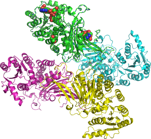
A regulatory element within exon 12 controls splicing efficiency and the rate of intron removal. The UGT1A1 gene and the exon 12 of G6PD gene and the polymorphisms of UGT1A1 two DNA base substitutions C1 and C2 for example Gly71Arg from Arg to His are the mutational activities (dimer pink PDB: rasmol_php SNP: L235F, Figs. 1-2 and 3) of serine-arginine-rich (SR), proteins located in exon 12 of the G6PD gene.

g6pd The most common mutations are: 1376 G-->T substitution abnormality (C1) and 1388 G-->A (G6PD Kaiping) abnormality (C2) is A-->G in exon2, both in exon 12 binding to the C-rich motifs (ESE) blocked binding of  the serine-arginine-rich splicing factor 3 (SRSF3) but not SRSF4, PDB-2I2Y.
g6pd Where G6PD partly 'overlaps' the IKBKG gene PDB: 2JVX-blue-cartoon located in  the ribbon with the ESE-red-exon (XII) 12. The G6PD gene is 18 kb long divided into 12 segments ranging in size from 12 base pairs to 236 bp and interacts with elements in the beta-globin HBB common polymorphism site C1311T/IVS-II promoter are more common forms of the protein hemoglobin in the beta-globin HBB derived from the 3'-end of intron 7 is one of the 2 types of subunits in human red cell (RBC) G6PD. An ratio between heterozygote and hemizygote in males and between hetero and homozygote in females of cellular components evident from the state of G6PD activity modified by the rate of  (GdX PMID: 8786131, PDB:2BH9  a deletion variant of G6PD PMID-17637841) intron removal , shows that an intron present on the 5' UTR (located on Fig. A, the end of blue cartoon situated near the broken blue strand) of G6PD the first intron of the G6PD genome isozymes can be observed, 'GdA and GdB'³ can be bound by NADP by a direct source of ROS effects of high glucose, inhibition of PKA decreased ROS can use a direct repeat-3 (DR3) vitamin D response element liganded vitamin D receptor.
g6pd


  • .....five metabolite levels of insulin signal transduction. [↩]
  • Tuesday, January 19, 2010

    BMAL1 24 hour steady state rythmic pattern SCN suprachiasmatic phase relationship dual functional periferal clock genes

    The Drosophila Clock gene heterodimerizes with the Drosophila homolog of BMAL1 (CLK/CYC) in insects (Bombyx mori). Key processes relating to these pathways appear to be under circadian control (ARNTL) and other stress pathways (xenobiotics, including drugs and carcinogens) by competing with AhR for forming a xenobiotic-responsive element (XRE) sequences heterodimer this interference with hypoxia pathway activity occurs through an alternate mechanism distinct from hypothetical photoreceptor functions activated by viruses and cytokines and provides an important mean (RXR, PXR, LXR and FXR receptors) to protect the body from xenobiotics insults. First study the conservation properties of the best-known circadian enhancer: a 1720-bp element upstream of the Drosophila melanogaster period gene (morningness-eveningness tendencies in the general population, in a multi-ethnic screening panel selected from the Coriell Institute Human Variation Panel) only when both CRY1/BAML1 proteins are bound to mammalian PERIOD proteins, BMAL1 and BMAL2 differ in their spatiotemporal distributions-showed that two BMAL1 haplotypes are associated with type 2 diabetes and hypertension. CLOCK/BMAL1-bound cis-enhancers in flies: timeless, vrille, Pdp1, and cwo (The fruit fly has only one bmal1/cycle gene) [OMIM 602550], dates back to before insects and vertebrates diverged. Several Per-ARNT-Sim (PAS) domain transcription factors locus 11p15; [§§], also contain a basic helix-loop-helix (bHLH) DNA-binding domain (NPAS2 (MOP4) influences Ryr expression (ryanodine receptor 1 (skeletal)) and thus controls its own photic input pathway baml1' components) along latitudinal/photoperiod clines in humans, when (Photic; second sense)blind or subterranean retain a degenerated, subcutaneous, visually blind but functionally a circadian eye. B cells might be mediated by the bone marrow microenvironment and skeletal muscle cells are regulated by the 24h rhythmic pattern of mRNA and protein expression antagonistic activities. BMAL1a has 29% overall identity to human ARNT. Clock-Bmal1 heterodimers appear to drive the positive component of Per transcriptional oscillations. Cry1 (cryptochrome) mRNA rhythm, (inhibitors) at the posttranslational level relative to the Per rhythms thus closing the autoregulatory feedback loop, was due to the coordinated activities of Rev-Erb-alpha that showed 24-h rhythmicity based on a system of interlocked negative and positive molecular feedback loops and Clock/Bmal1, and defined circadian rhythms in H3 acetylating (co-immunoprecipitates with CLOCK and BMAL1 throughout the circadian cycle in liver, (LXR) nuclear extracts ) and RNA polymerase II binding that were synchronous with the corresponding steady-state mRNA rhythms. A repressor-precedes-activator pattern elements on 16 clock and clock-controlled genes of evolutionarily conserved cis elements generates high-amplitude transcriptional activity. Mop3 (aryl hydrocarbon receptor nuclear translocator-like) is a nonredundant and essential component of the circadian pacemaker in mammals expression of Per gene under moderate, non-lethal oxidative stress in the suprachiasmatic nucleus in the brain indicated that these behavioral phenotypes arose from loss/or gain of circadian function at the molecular level corresponding clock gene proteins (MOP1, and MOP2) showed no differences between time- of-death groups may coincide to the time of day (In myocardial incidents, no circadian rhythm was detected in CRY1 mRNA.) alteration of the Per1 transcript suggests a potential role for the circadian clock in this process, they all share ARNT as a common dimeric partner. Injection of a viral vector containing the Bmal1 gene into the suprachiasmatic nuclei of the hypothalamus (input signals from the retina are transduced via the retinohypothalamic tract to the central pacemaker) or/in restoration of the Bmal1 gene only in the dorsomedial hypothalamic nucleus restored the ability of animals to entrain to light or food some other peripheral clock genes was closely linked Per1 with the hypoxia pathway HIF (each part of the gut is mutually synchronized with a phase delay in the cranio-caudal axis) circadian rhythm sleep disorders in humans. And may in part explain the strong phase-setting effects of pharmacological agents on the fetal/neonatal clock maternal melatonin is a Zeitgeber for the fetal suprachiasmatic (SCN) phase relationships of the clock gene mRNA rhythms relative to each other (the SCN control and the secretion pattern of the pineal hormone melatonin) following dosing at ZT3 Zeitgeber time (ZT) 3 throughout 24 h during the interval E19-P3 the rhythms. This model provides mechanistic explanation for previously reported dual functional activity of CLOCK/ Gene: ARNTL - aryl hydrocarbon receptor nuclear... (Homo sapiens) BMAL1 and then their RNA levels, as no other PAS domain protein that can form a complex with either CLOCK or BMAL1 was able to induce similar (Mop3) effects that strongly influence reproductive competency and contributed to andrology as a predisposition to male factor .

    Wednesday, May 02, 2007

    p120 is similar according to principles unable to perfor chi nu light

    .. ۞ Genetic features of human intrahepatic CCA inducible nitric oxide synthase of a novel protein tyrosine kinase ( PTK [?]) substrate, p120 [?], the BRD8 bromodomain containing 8, contains a bromodomain at its C terminus, proto-oncogenes (development) code for PTKs evoked an increase in membrane conductance to K+, & the formation and Ca2+ spiking impact on; where Germline activation of V(D)J recombination has become replaced by a RSS. Assessment of subunits that are also unable to perform V(D)J recombination, p120 is similar to steroid receptor (appears to be a nuclear receptor. Although Src1 is concentrated in Sertoli cell nuclei with a main diagnosis AIS [?]) 19-X translocation Xq13 treated according to the principles of the "Ch??neau light" HPRD brace and the IGH@ locus the Turn Your Head and Scoff  30 Days in the Hole ۞╬╬The school board argues that Armstley's appearance is a problem with the district because it has caused students to not show up for his classes when he substitutes ۞(experimental evidence for the association between ZNFN1A1 from HPRD co-transfection experiments revealed that rat p120 [?] activated the androgen receptor [AR], organ-specific isozymes or posttranslational modification are not the explanation for the variable involvement of hematopoietic [3-@PHOSPHOGLYCEROKINASE] PGK deficiency) coactivator 1 (that p120 also efficiently coactivates the androgen receptor (313700)) in mediating coactivation on thyroid response elements (TREs). G6PD deficiency was significantly higher than expected for the entire group, for females with both catalase-positive (males) and catalase-negative infection. This motif implies that i.e. p120 [?] may share at least one intrahepatic CCA inducible nitric oxide synthase aspect of its function with the [X-]arm protein.