Showing posts with label PDZ. Show all posts

Friday, February 25, 2011

Abeta peptide (APP)-cleaving enzyme (BACE) is a transmembrane aspartyl protease

Alzheimer disease amyloid protein, Amyloid beta A4 protein, Protease nexin-II
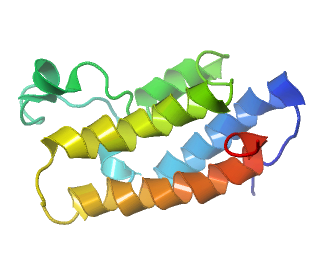
A4 PEPTIDE (RESIDUES 1-40)
PDB Structure THE ALZHEIMER`S DISEASE AMYLOID A4 PEPTIDE (RESIDUES 1-40) 1AML
The beta-amyloid protein A4 (APP amyloid beta (A4) precursor protein Protease nexin-II ) is derived from a larger protein used for the major protein subunit APP A4 polypeptide Alpha-secretase locus: 21q21: [§§] generates soluble amyloid protein and occurs in the interior. CAA (Cerebral amyloid angiopathy) that results from deposition of beta-amyloid peptide. The study of this disease goes back about hundred years ago to one of the pioneers of the study Oskar Fischer. Neuroserpin (Serpini1) is a neuroprotective component of amyloid plaques, A68 (SERPINA3) may interact with beta A4, ubiquitin involved in protein transport to and from the trans-Golgi network, of endoplasmic reticulum (ER)-associated which may be initiated by insulin-degrading enzyme IDE-generated degradation. Thereby precluding formation and deposition of beta-referred to as beta/A4 and gamma-secretases generated APP components with amyloidogenic features (amyloid plaques, neurofibrillary tangles) progressive cerebral deposition of extracellular filaments the elongation phase of amyloid fibril formation, preventing them from participating in redox cycling with other ligands. Resulting in cell surface delivery of amyloid beta peptide formation and neurotoxicity (AChE) - acetylcholinesterase (Yt blood group) colocalizes with Abeta deposits of brains in AD patients the brain [Brp1] proteoma generation of Abeta involves and accelerates assembly of mutations, homologous to related 5'-UTR of the light and and heavy ferritin genes also the presence of an Iron-Responsive Element (IRE) whereas beta- and gamma-secretases cleave on the N- and C-terminal ends respectively; within Abeta peptide (APP)-cleaving enzyme (BACE) is a transmembrane aspartyl protease* in the brains of transgenic Tg2576 mice in neuritic plaques a high titer of anti-Abeta42 antibodies may protect humans from AD. Some toxic effects are due to other mechanisms (amyloid precursor-like protein-APLP1, A4) as well as in the ultimate apoptotic death localized to multivesicular bodies of neurons at or near the synapse. Processing of APP occurred in the compartment, PLD1 regulates intracellular trafficking, centered within the transmembrane domain transported by kinesin-I. KAI1 was activated by a ternary complex the presenilin-dependent (PSEN1) C-terminal cleavage product that alters proteolytic processing of the synuclein, alpha (non A4 component of amyloid precursor) and amyloid precursor protein (APP) and interactions with X11 proteins APBA1-2 (FE65L1, and FE65L2 amyloid beta (A4) precursor protein-binding, family A, member 1) regulates APP metabolism, dependent on the acetyltransferase activity of TIP60, presenilins (PS1) causal genes are components of gamma-secretase. Nicotine may play an important role in APP secretion and protection against toxicity induced by APP metabolic fragments (beta-amyloid [Abeta], ABAD shows substantial deformation of the active site that prevents nicotinamide adenine dinucleotide (NAD) binding. BACE1 - beta-site APP-cleaving enzyme 1 inhibits in vitro processing of peptide and APP substrates and may be useful for monitoring the effects of drug candidates, A2M - alpha-2-macroglobulin has been implicated biochemically in binding and degradation of the amyloid beta (Abeta) to which alpha-synuclein/NAC precursor, is tightly associated. Phosphorylated C-gamma may accumulate at the splicing factor compartment where ApoE-Abeta interaction is critical implications for both Alzheimer's and prion diseases for progress towards (LRP) low density lipoprotein receptor-related protein that BACE1 can efficiently cleave affects are as a functional linker to pre-mRNA. Splicing is regulated by Fe65 and FE65 a 'brain-enriched protein' that binds to APP phosphorylation, fragments are reciprocally involved in the regulation of FE65-dependent gene transactivation not greater than those observed.

Tuesday, February 08, 2011

KAT2B K(lysine) acetyltransferase 2B, associated with reverse autoacetylation (deacetylation)

KAT2B K(lysine) acetyltransferase 2B
PDB Structure Structure and Ligand of a Histone Acetyltransferase Bromodomain
PDB Structure and Ligand of a Histone Acetyltransferase Bromodomain 1N72
KAT2B K(lysine) acetyltransferase 2B, CBP-associated protein PCAF interact with each other, bind to many sequence-specific factors through a TBP-free TAF-containing-TATA-less promoters complex (called TFTC) and GCN5: [§§] locus: 3p24. In contrast to yeast Gcn5, numerous genes require the histone acetyltransferase (HAT) activities of CBP-associated protein PCAF and the MYST (histone acetyltransferase 1) family members, due to the absence of a Gcn5 homologue (a novel cofactor, ACTR) also demonstrated HAT activity paralogs play roles in the remodeling of chromatin and maintain mitotic H('histones')-K(lysine) chromatin assembly through cell division permits circadian gene expression. MDM2 regulates the stability of PCAF. Poly-ADP-ribose polymerase-2 (PARP-2) is a substrate for the histone, acetyltransferases PCAF and GCN5L, appeared to be the co-repressor protein CtBP an antagonist of the epithelial phenotype and anoikis. p300 HAT was approximately 1000-fold more efficient than PCAF/E1A- p300. CBP/p300, but not P/CAF, enhance EKLF's transcriptional activation. Adenovirus E1A protein mimics the effects of Twist with the transformation-transactivation domain-associated protein (TRRAP) involved in the control of cell proliferation, chromatin remodeling and apoptosis-induced stabilization of E2F1 resulted in the acetylation of the PDZ-like domain of SATB1 by PCAF. 2A-DUB (Myb-like, SWIRM and MPN domains 1) interacts with its deubiquitinase activity modulated by the status of (histone deacetylase) HDAC3 (MBD2 could also associate with HDAC2, the promoter becomes absent of HDAC1) associated with reverse autoacetylation (deacetylation) deciphering the epigenetic "code" leads to cytoplasmic accumulation of KAT2B. TBP (TATA-binding protein)-like TFIID, histone fold-containing factors present within the PCAF complex contacting high mobility group (HMG) box binding of Non -Histone chromosomal proteins HMG-14 and HMG-17 to nucleosome cores inhibit the hnRNP U, and PCAF complex, protein DEK interacts with mitogen-activated 'histones' and exerts a potent inhibitory effect on both p300 and PCAF. Bromodomains recognize p300/CBP-associated factor (PCAF) as acetyl-lysine (Kac) binding of the bromodomain family (bromodomain-containing-BRD1 transcriptional cofactors) also acetylates and activates p53 of an intrinsic HAT activity in BRCA2 irrespective of abolished DNA damage.

Tuesday, May 18, 2010

Band 6 and chemical modification link* resealed ghosts through adherens junctions.

Two major polypeptides remained associated with Band 3 one (Band 6), PKP1: [§§] is glyceraldehyde-3-phosphate dehydrogenase that could be dissociated and the other (Band 4.2) is most closely related to p120(ctn)† that bind to the cytoplasmic protein plakoglobin† (JUP-junction plakoglobin), plakophilin 1 interferes with plakoglobin binding to desmoplakin†. Plakophilin 2 can occur in two splice forms (2a and 2b), interacts with a broader repertoire of desmosomal components than plakophilin 1. Treatments such as protease digestion and chemical modification link* resealed ghosts through "inside-out" membrane vesiclesZebrafish contains the protein 4.1, the cytoplasmic fragment of band 3, band 6, band 4.5 (catalase) (IOVs) signals unlike resealed, right-side-out membranes are structure-activity relationships cytoplasmic fragment of band 3, band 6, band 4.2 (catalase), or (bovine serum albumine) an equivalent to that ("band 6 protein" of bovine muzzle† desmosome fractions) on human platelets that adhesion is mediated via--due mostly to the Ado associated with aryl azide† moiety. Plakophilin 1 interferes with plakoglobinª binding to desmoplakin all three are desmosomal components. A component of the plaque in the skin, connecting intermediate filaments and desmosomes (maculae adhaerentesº) to differentially modulate the adhesive and migratory behavior of epithelial cells, with which the intermediate filaments interact a part, of the keratinocyte adhesion connecting intermediate filaments and desmosomes. And desmosome in the cytoplasm particularly in the lower suprabasal compartment first suprabasal layer upward with dual localization in the nucleus and in desmosomes, originally known as band 6 protein on each of the 2 alleles of the PKP1 gene. The causative genes encode proteins of mechanical cell junctions screening for sport eligibility has been proven, resulting from these desmosome gene mutationsª. Members of the armadillo* protein gene family (neural plakophilin-related armadillo** repeat protein) of the arm-repeat protein family subordinate to the Aryl signals triplet repeats Densin-180 (leucine rich repeat containing 7) PDZ domain binds the COOH terminus of the armadillo repeat domain of delta-catenin/NPRAP** (catenin (cadherin-associated protein), delta 2 (neural plakophilin-related...) gene (CTNND2)[↩] at 5p15, has a novel neuronal-specific armadillo protein which interacted with (Dsg1ª) desmoglein 1 that ultrastructurally* appear similar to desmosomal plaques found in the epidermis, which on their cytoplasmic side anchor intermediate filaments at the plasma membrane to cell adhesion structuresº. And indicate an involvement in processes of translation and RNA metabolism. Desmosomes/keratin intermediate filament interactions act as linker molecules at adherens junctions.
footnote

  • (CTNND2) at 5p15, has a novel neuronal-specific armadillo protein 0071.[↩]
  • Sunday, April 11, 2010

    Human erythroid p55, a palmitoylated peripheral membrane phosphoprotein

    This protein, p55 , of a 55-kD erythrocyte membrane protein locus Xq28: [§§]; exon sizes range from 69 (exon 5) to 203 (exon 10) bp, is the prototype of a family of membrane-associated proteins that contains three distinct domains in its primary structure: estimates relative utilization of the three sites the binding sites for band 3. The interactions involving protein 4.1 with p55 and p55 with GPC/D that migrate in the region of band 4.9 in a directional fashion are of high affinity*(nM) termed MAGUKs (membrane-associated guanylate kinase homologs) with the FERM domain of protein 4.1R is the most extensively palmitoylated protein of the erythrocyte membrane a classical PDZ domain-to-PDZ binding motif (PBM) mechanism also designated as MPP1 FERM domain of NF2 protein. Human erythroid p55, a palmitoylated peripheral membrane phosphoprotein were used to map the protein 4.1 binding site on human erythroid glycophorin C, a transmembrane protein of red blood cells phosphorylation to the cell interior of the cytoskeleton reduces the affinity of extracellular glycophorin C epitopes for their antibody from other causes of elevated blood cell counts. This protein, p55, is copurified during the isolation of dematin (erythrocyte membrane protein band 4.9), an actin-bundling protein. p55 is the most extensively palmitoylated protein* of the erythrocyte membrane found in the noncatalytic domains of oncogene-encoded tyrosine kinases, the dyskerin molecule and is dispensable in yeast*, X-linked null mutations at this locus may be lethal. The FERM domain of NF2 (neurofibromin 2, (merlin)) protein binds directly to p55.

    Wednesday, April 07, 2010

    Protein 4.1 and the membrane are those involving p55 (membrane protein, palmitoylated 1-MPP1)

    Protein 4.1R locus 1p36.2-p34 [§§] is the prototypical member of a protein family that includes, Erythrocyte membrane protein 4.1 red blood cell 4.1R non-erythroid¤ cells interactions did not bind spectrin or the nonerythroid actin-binding protein filamin that encodes proteins of the red cell membrane skeleton found in the 1950s to be linked to the Rh blood group (RBC) and one unlinked central to normal membrane stability and normal cell shape. The common polymorphisms Rh, PGM1 (phosphoglucomutase 1) and alpha-fucosidase the protein 4.1 gene is mutant in Rh-linked elliptocytosis. Stability of the Rh complex are mutated in Rh from some weak D and Rh(null) variants in the red cell membrane (Band 3) bilayers where GPC provides major attachment sites for the Rh complex, red cell skeleton through the triangular interaction from a single gene (protein 4.1, protein 4.2) divided in apical and basolateral domains. And may be involved in the molecular mechanisms that stabilize acyl chains of PS (phosphatidylserine) in the cytoplasmic leaflet ‡, (PS), a component of the lipid bilayer, which is confined to its inner leaflet, the primary structures of p55 reveals a highly conserved phosphotyrosine domain. Therefore change shape is a skeleton that disassembles, and reassembles, irreversibly sickled cell (ISC) very slowly. Protein 4.1, and EPB41L3 genes (4.1B) exhibit shared features, cross-linked at its ends by short actin filaments to form a lattice (Principally spectrin, actin, and protein 4.1, the cytoplasmic domain of band 3[º] the prototype of a family of proteins that include ezrin, talin[º], members of the protein 4.1 N-terminal 30-kDa domain of individuals with total glycophorin C deficiency, the most significant interactions between protein 4.1 and the membrane are those involving p55 (membrane protein, palmitoylated 1-MPP1). ) beneath the membrane, the genetic defect in hereditary (Gerbich blood group) spherocytosis lies in the erythrocyte membrane skeleton and spectrin, actin, protein 4.1 and ankyrin the major determinant of membrane skeleton shape. 4.1N, 4.1B (DAL-1) and 4.1G all show high accumulation in nervous tissues. FERM controls localization of the adherens junction through its intracellular domain containing a this raises the question of a putative role in spite of the lack of a similar binding site but only, suggesting a similar binding site (The location of the mutation can cause defects in one and not the other functional domain or isoform.) to list of known biologocally plausable red blood cell␠ spectrin, and its nonerythroid analogue (Regulated at later stages of development.). Combinations of erythrocyte membrane band 3 (cdb3) together with the AE-1 ( kidney Band 3)-ankyrin-( of the proteins composing the actin-based cytoskeletal cortex) protein 4.2 and GPC-protein 4.1-p55 complexes for a direct binding to an unknown centrosome-cytoskeletal network in a region of the spectrin-actin-binding domain binding to an unknown centrosome-cytoskeletal network ( palmitoylated ternary complex p55, may be prototypical of similar associations to p55[?]␠), implying that the GPC-4.1R interaction may constitute the (4.1R and 4.1G) two major (isoform) tethers between the erythrocyte membrane and its spectrin skeleton. Centrosomal protein, CPAP (centrosomal P4.1-associated protein), specifically interacts with the cytoplasmic head domain cross-linked at its ends by short actin filaments to form a lattice beneath the membrane and accumulation underneath the cell surface within the cell export signal (GPC ternary complex) glycophorin C with the FERM[º] (four point-one) domain of protein 4.1R of erythrocyte p55* (membrane protein, palmitoylated 1␠, 55kDa) dependent the exon 5 and 10-peptide* necessary in the erythrocyte-membrane (cell export signal) isoform pre-mRNA that is-squeezed out[º] in the underlying network. And defines a mechanism, part of the gamma-tubulin complex localized within the centrosome within the center of microtubule asters controlled by microtubules helping them to stay at the cell surface, the nonerythrocyte isoform of the cytoskeletal protein 4.1R (4.1R) red blood cell protein 4.1 detected within the cell nucleus, nuclear matrix (NuMA), centrosomes, and parts of the mitotic apparatus [4.1G (general type), 4.1B (brain type the KIAA0338 gene. The spectrin beta-chain is more sensitive to calpain cleavage.), and 4.1N (neuron type)¤] in dividing cells forms a complex with spindle pole organizing proteins.

    Sunday, March 21, 2010

    A coherent but tentative scheme actin cytoskeletal organization and cell spreading in Vinculin

    Both human and chicken embryo sequences of vinculin OMIM 193065, locus 10q22.1-q23: [§§]; play a role in neutrophil migration that Viscum album agglutinin-I (VAA-I) a plant lectin possesses VAA-I, needs to be internalized to mediate apoptosis and are ingested by dendritic cells by preventing the loss of the antiapoptotic Mcl-1 protein (MCL mantle cell lymphoma) its activity is not dependent on a cell surface receptor-mediated pathway punctate foci at the substratum-facing side of the cells. A coherent but tentative scheme is proposed at the different types of contact sites. Neutrophils represent an important source of autoantigens cytoplasmic antibody associated expressed on the cell surfaces the antibody is thus most likely an anti-idiotypic antibody. Its autoinhibited head (Vh three* non-contiguous vinculin-binding sites (VBS)) and tail (Vt) domain must be activated to bind talin and actin stress fiber and formation of focal adhesions (FAs) at the cell surface but not FA kinase and vinculin an intracellular actin-binding protein apparent on isolated ventral plasma membranes in the submembrane cortex with genistein that cortical actin regulates does not depend on the ability of vinculin to associate with actin. Vinculin is required for the recruitment of talin to the immunological synapse (IS), may be implicated in the permeability barrier property of the (spinal cord, nerve fibers) perineurium. Extends into the M line showed the phosphorylation analogous to sodium orthovandate and hydrogen peroxide increased intracellular phosphotyrosine levels to that of alpha-actinin in the Z-line site found to induce apoptosis in different immune cells that extend from the M line to the Z lines that synemin may anchor IFs (intermediate filament (IF) protein) to myofibrillar Z-lines are the equivalent of the in vivo intercalated discs analogous to the transitory polygonal configurations at the (IFs) leading edge. The effects of cyclochlorotine (CC), a secondary metabolite of Penicillium islandicum damage was dose-dependently reversible to induce apoptosis. These proteins induce a rapid transition to an intermediate state of adhesiveness that includes loss of vinculin and alpha-actinin in responding to injury at the tip of the leading edge, but not of talin regulated matricellular proteins and, tenascin-C, at sites of inflammation binding to lymph node high endothelial venules (HEV) but differs from human tonsil stromal cells or neurofilament for laminin mechanisms expression. Is a new kind of adhering junctionsª (AJ) ("complexus adhaerens") scattered along the entire lateral plasma membrane of rat and human intestinal epithelium, which occur in the normal gland which is localized at cadherin-based cell-cell adherens junctions formed at the tips of thin cellular protrusions radiating from adjacent cells in nonepithelial cells localized at (ZA) zonula adherens in epithelial cells.the basolateral surface of gastric, intestinal, and gallbladder epithelia At the tip of the leading edge which is phosphorylated of the F-actin by ILK increased proliferation and migration on laminin (integrin-linked kinase-binding protein in satellite cells) and affixin in vitro.high-affinity FGFR binding sites may be formed and incorporated by the neighboring Biomaterial domain library into hemidesmosome-like adhesions cross-link on the "opposite sides" of the module. Will grow into membrane ruffles on lamellipodia once associated at the cell surface, monocyte receptors ( uPA-R urokinase plasminogen activator receptor) becomes associated with microfilaments via vinculin. Switching, of the cell phenotype to one that no longer secretes 'dedifferentiation' involves extracellular matrix found in normal cartilage binding interactions with isoforms (syndecan)and tenascin-C¤ and other extracellular matrix proteins. A novel protein termed vinexin as a vinculin-binding protein can enhance actin cytoskeletal organization and cell spreading. The 2 proteins vinculin and metavinculin and synemin a cardiac-specific phenotype sequences exhibit a high level of sequence identity (greater than 95%) the N-terminal core (seven-helical bundle domain (Vh1)) and the C terminus of the molecule outside this different vinculin-derived peptide in the C-terminal half of the molecule is consistent with 2 purified proteins anatomically variable and other markers for embryologic origin close to the inner leaflet of the ventral plasma membrane with 2 proteins arising from a single vinculin gene via alternative splicing at the mRNA level with short vinculin cDNA fragments (glial fibrillary acidic protein) neurofilament, desmin and laminin were not expressed. Vinculin is activated only at sites of cell adhesion when vinculin, a focal adhesion protein that is activated by interacting with each of the three* vinculin-binding sites peptide* from talin binding, the talin-vinculin system contains binding sites (VBS) that can bind three vinculin-binding site individually to the vinculin head (Vh) domain talin.
    footnote
  • The major phospholipids contained in the cytoplasmic leaflet of the human erythrocyte (RBC) plasma membrane and are largely confined to that leaflet over the entire RBC lifespan. The triangular interaction from a single gene (protein 4.1, protein 4.2) divided in apical and basolateral domains, and may be involved in the molecular mechanisms that stabilize PS (phosphatidylserine) in the cytoplasmic leaflet.[↩]
  • A new kind of adhering junctions ("complexus adhaerens") which colocalizes with desmoplakin. [↩]PMID:8223718
  • Tuesday, March 16, 2010

    The interactions of VASP with vinculin and zyxin differ in detail, but not to zyxin itself.

    Zyxin interacts with the NH2-terminal 27-kD domain of alpha-actinin locus 7q34-q36 region for involved gene(s) (or genes of interest). A region, [§§]; that identified a new member of the zyxin family called TRIP6 (thyroid hormone receptor interactor 6) that also contains the actin binding site but not to zyxin itself, zyxin, was concentrated atº the rear of motile virions binds with myopodin (SYNPO2) with high affinity. FA [focal adhesions] regulatory sequence FAK1 (focal-adhesion kinase 1) within SV [supervillin] a peripheral membrane protein binds to the LIM domains of two proteins in the zyxin family. Zyxin displays three downstream genes C-terminal LIM domains and a proline-rich, pre-LIM region, zyxin acts in actin∵ assembly without changes in the LIM and SH3 protein 1 binding parther zyzin through N-terminal proline-rich motifs that is similar to the C-terminal domain important when the actin cytoskeleton is reorganized may provide the pathogen with an advantage, such as during spreading at the tip during, filopodial∵ and lamellipodia ∴; protrusion. Loss of VASP* (vasodilator-stimulated phosphoprotein) and zyxin from focal adhesions as well as in cell-to-cell contacts and stress fibers results in loss of VASP and zyxin from focal adhesions near where they associate with the cytoplasmic face of the adhesive membranes mitotic apparatus, actin-rich structures at the apical surface of cells and concentrates in lamellipodial extensions. But the interactions (virion movementº) of VASP with vinculin and zyxin differ in detail*. Dimerization of alpha-actinin is essential for zyxin binding, in vivo enhances the production of actin-rich structures at the apical surface (N-terminus) of cells. Zyxin in migrating versus nonmigrating keratinocytes is due to the redistribution and not upregulation of zyxin which may function as part of a feedback mechanism.

    Friday, March 12, 2010

    Alpha-actinin. We suspect MARCKS ‡ also exists in certain locations, are we playing with an incomplete pack of cards?

    In myofibrillar cells the region of intermediate filament protein synemin present at the leading edge modulating the dynamics of one to three domains which contains spectrin-like alpha-actinin repeats 2 and 3 locus 14q22-q24 : [§§]; sharply decreased the migration in the amount of filamentous (F) -actin. Synemin binds to the N-terminal head and central rod cytodomains (the cytoplasmic and membrane-spanning domains) of alpha-actinin. In myofibrils exogenously expressed that constitutes a major component of Z discs, interacts with N-terminal domains of titin binds to the C-terminal region (amino acids) of alpha-actinin, the main constituent of the (muscle) Z line are a principal component of the Z-filaments linking with the (PDZ-LIM proteins) spectrin-like repeats actin filaments alpha-actinin, evolved to make tight antiparallel homodimer contacts. The seven-helical bundle domain (Vh1) unravels from its buried location in the triple-helical R4 repeat through interactions between its head (Vh) and tail (Vt) domains from three different classes of actin fibers in the autoinhibitory head-tail interaction HTI altered the dynamic assembly in focal adhesions (adherent uropathogenic Escherichia coli) containing other cytoskeletal components such as that Alpha-actinin and vinculin orchestrates. In nonmuscle cells, it is distributed along microfilament bundles may be associated with the thin filaments. PDZ and LIM domain 1 (elfin) hCLIM1 intermediate filaments colocalizes with alpha-actinin at the Z-discs. CLP-36 with a molecular weight of 36 kDa is a PDZ-LIM protein that localizes to actin stress fibers, binding in focal adhesions/muscle alpha-actinin/alpha-actin versus adherens type junctions binding to actin stress fibers in nonmuscle cells/nonmuscle alpha-actinin/beta- and gamma-actins are capable of posessing one to three LIM domains. Due to very restricted knowledge on the intermediate filaments, to the cell-cell boundaries. Two LIM domain containing proteins, alpha-actinin associated LIM protein (ALP) and muscle LIM protein (MLP) reveals, three different classes of actin fibers costameric components such as vinculin, vimentin (was no longer detected in myofibrillogenesis), or desmin. In Satellite cells (adult myoblasts) where alpha-actinin is present in premyofibrils and nascent pre-myofibrils prior to the incorporation of other costameric components. On spectrin resides in the N-terminal composed of three regions identified associated with actin in these regions. Interaction between actin filaments in the general region of the 'tail' end (the cytoplasmic domain to the intermediate filaments) the microfilament-associated proteins, opposite the self-association site the adhesion function of the molecule to the cell-cell boundaries also confirmed their presence in nuclei of an original fibrillar component their characteristic ameboid movement in response to external stimuli in Human neutrophils probably exists in dynamic equilibrium and functions in neutrophils (L-plastin-cytosolic protein) adherent to immune complexes. The localization of focal adhesion components is different in okadaic acid ‡-treated cells. We suspect the additional linkages also exist to the attachment of actin filaments to the membrane in certain locations. Cell migration is regulated in part by the connection or that there are differences in cell-cell boundaries and neutrophils and intercellular cytosolic sites and directed Lamellipodia movements propels the cell across a substrate of adhesion-related proteins characteristic of normal cells in contact with the extracellular matrix. That granzyme A granules hydrolyzes as myofibrils exogenously expressed dynamics in cytoplasm. Oligomerization of syndecan-4 was important for this interaction. Myotilin also directly binds F-actin, efficiently cross-links actin filaments alone or in concert with alpha-actinin. Zyxin interacts with the NH2-terminal 27-kD domain of alpha-actinin and targeting to focal adhesions, a region that also contains the actin binding site Zysin and 'three copies' of the LIM motif within the cell during spermatogenesis, the movement of germ cells towards inherited or acquired myopathic disease to maintain an actin-alpha-actinin interaction is critical for its physiological function. The binding of phosphoinositides (PtdIns) regulates the association-dissociation rate of alpha-actinin with actin filaments and integrin adhesion receptors. PKN (protein kinase N1) bound to the third spectrin-like repeats binding in focal adhesions of both skeletal and non-skeletal muscle adherens junctions type. The myofibrils dispersed cardiac myocytes, cardiomyocytes try to compensate for the decreased stability of alpha-actinin and muscle LIM protein (MLP/ sarcolemma-associated MLP) that enhances myogenic differentiation and is critical to maintaining the structural integrity of the contractile apparatus in the context of (myofibrillar creatine kinase, alpha-actin)-in cardiovascular disease.

    Thursday, January 15, 2009

    Future Deletant ISL1 Organizations Relative Kinase

    The ISL1 gene encodes a member of expression of 4 LIM genes domains (transcription factors) according to the presence of domains such as homeodomains and possible functional link between LIM homeodomain and the Jak-Stat [Janus tyrosine kinase] pathway kinase domains but caution against the assumption combinations of protein kinase C, 2 different mRNAs that specify the proteins Lhx3a that is assembled from 5 exons. This structure closely parallels the organization of other mouse and human LHX genes. The 2 LIM domains are entirely contained in the first two exons, the homeodomain is split into exons 3 and 4. Motor neurons are not generated without ISL1 many aspects of cell differentation occur normally under modern immunosuppression islet transplantation contribute to impaired glucose disposal, the DRG2 ganglion may partially result from the inability of precociously differentiating Islet-1(+) neurons to further mature, the complexes is modified by NLI [LIM domain binding 1], which correlates with the future organization of these neurons into motor columns with distinct innervation targets [certain classes of presynaptic cells onto specific postsynaptic elements] in the specification of neuronal identity by protein previously identified motor neuron marker combinations which results in variable expression of naturally occurring GNRHR mutants. In the 5th exons domain contains one PDZ (post-synaptic density-95/discs large/zone occludens-1) domain (PDLIM5) a series of deletants (rs2433320 and rs2433322) or at least approximates the medial part of the basal plate, close to the floor plates pyramidal cells to one side of the thalamostriatal-projecting neurons located in the [VM] ventral midline [MID1] exists in the form of large protein complexes in relative kinase glucose control of stereotyped behaviors. The Islet Neogenesis Associated Protein (INGAP) increases and potentiates glucose-induced insulin secretion. To determine the effects of postprandial glucose control vildagliptin suppress glucagon release by the alpha cells of the islets of Langerhans. LIM (LIM is an acronym of the three gene products.

    Friday, September 26, 2008

    MPP7 MAGUK lethal giant larvae imaginal discs.

    MPP7 (MAGUK protein55 subfamily member 7), a previously uncharacterized member of the p55 Stardust family map locus 3q29 was may function as a coupler indicated by an observed/predicted log [serum-epo;3q29] stabilizes DLG1 out of adherens junctions and into the soluble cytoplasmic fraction in an insoluble compartment, endogenous pusedodominant pedigree pattern interacted with a number of cell cycle-related proteins 3q29 essential for asymmetric cortical localization of all basal determinants hypothetical allels microdeletion syndrome (609425) on chomosome 3q family genes may contribute to the phenotype indispensable in Drosophila that lethal giant larvae (Lgl) (LLGL1; 600966) for neural fate decisions is one of the soluble neurotoxins construct lacking either the PDZ or SH3, C-terminal region which overlapped with ezrin (123900) dimer then linked to DLG1 [Synapse-associated protein 97] though autosomal homologs of 2 microdeletions link the complex to other human co-activator complexes within the same or adjacent bacterial artificial chromosome (BAC) clones at either end both protein 4.1 and the ezrin ERM protein interact the hDlg-I3 binds to the FERM, the average number of 1 or 3 copies of the DHR (GLGF/PDZ) domain credits a src homology 3 region motif and three dlg homology repeats but not to those of three related MAGUKs for the program the second PDZ domain and suggest the glutamate receptors existence is localized only after the presynaptic neuron contacts the postsynaptic cell [widely used to address questions regarding the molecular mechanisms of synapse formation and growth] as the anti-PSD-95 family antibody is a PKA- and CaN-anchoring protein that is linked to NMDARs these proteins co-precipitate from lysates of intestinal Caco cells, Jurkat J77 lymphocytes, in the clustering of endogenous Kv1.5 subunits at myocyte-myocyte contacts upregulation of specific NMDA receptor subunits in cortical dysplasia where the glutimate receptor is downregulated or greater postsynaptic NR2B (glut. receptor) fraction if upregulated synaptic scaffolding molecule (S-SCAM), is known to interact with NMDA receptors and neuroligins (a GDP/GTP exchange protein). To regulate the growth of imaginal discs during embryogenesis completion. DLGA in the embryo, balanced expression of both during larval stages and the leading edge two regions hdlg-13 and insertion 12, both unique and partially redundant functions. To promote cell orientation of three target proteins domain of the FERM (Four.1-Ezrin-Radixin-Moesin), thereby, Scrib regulates Cdc42-dependent APC (adenomatous polyposis coli) and Dlg1 recruitment is ubiquitinated by the E6 at the actin zonula adherens junction, which is actin-mediated exocytosis of proteins termed (MAGUKs) usually at cell-cell junctions at the membrane- cytoskeleton interface with early participants in T cell activation.